CORNEAL ULCER IN A CAT
Today we bring to your attention an article on the topic: “corneal ulcer in a cat” from professionals for people. We tried to fully cover the topic. If something is not clear, then experts are ready to answer all questions in the comments.
A corneal ulcer is a defect on the outer layer of the cornea, which is caused by necrotic damage to the stroma and epithelium. The main danger of the disease is the rapid development of complications with perforation of the eyeball, the development of panophthalmitis and subsequent removal of the eye.
A cat’s eye is a delicate structure that can be damaged by external influences.
An ulcer can develop in 2 directions – infectious and non-infectious.
Non-infectious factors for the onset of the disease include:
- Injury.
- Breed predisposition – most often in brachycephalic cat breeds (Persian, exotic, Himalayan, etc.).
- Turning of the eyelids.
- Incorrect growth of eyelashes, which constantly injure the cornea when blinking.
- Entry of a foreign body.
- Dry eye syndrome.
- Decreased local immunity.
- Chemical burns to the eyes.
The infectious nature of the occurrence can be caused by both bacteria, viruses and fungi. Most often, the eye is subject to inflammation due to staphylococcal, streptococcal infections, Pseudomonas aeruginosa, corona and herpevirus, as well as chlamydia. This type of ulcer has a long and severe course, is prone to relapse and is difficult to treat.
A healthy cat’s eye has 4 layers of cornea. The outer layer is represented by multilayer epithelium, which mechanically protects the organ of vision of animals. Below is a kind of framework – the stroma, which consists of collagen fibers and has the greatest thickness among all layers of the cornea.
Under it there is a membrane, which is a “guard” against the penetration of harmful flora. The innermost layer is the endothelium; it is needed to maintain the water balance of the eye. The main function is to remove excess fluid to maintain the transparency of the entire cornea and prevent edema. Does not have the ability to recover from damage.
If only the outer epithelium is damaged, then the body copes with the injury on its own, “healing” the defect within a few hours. However, when a microorganism enters the eye tissue, toxins and waste products of pathogenic flora begin to be released. These substances deepen the lesion, melting the tissue and causing perforation of the cornea, as well as the introduction of infection into the deeper layers.
The disease varies in severity of symptoms, depending on the presence of the pathogenic agent and the presence of complications.
The main features include:
- At the initial stages, narrowing of the palpebral fissure, swelling, hyperemia and lacrimation occur.
- A sharp pain appears in the eye, the animal often shakes its head and scratches the damaged eye with its paw.
- Photophobia and constant squinting.
- Subsequently, the ulcer penetrates into deeper layers, which are not painful when damaged due to weak innervation, and the initial symptoms of inflammation disappear. Purulent or mucous discharge appears.
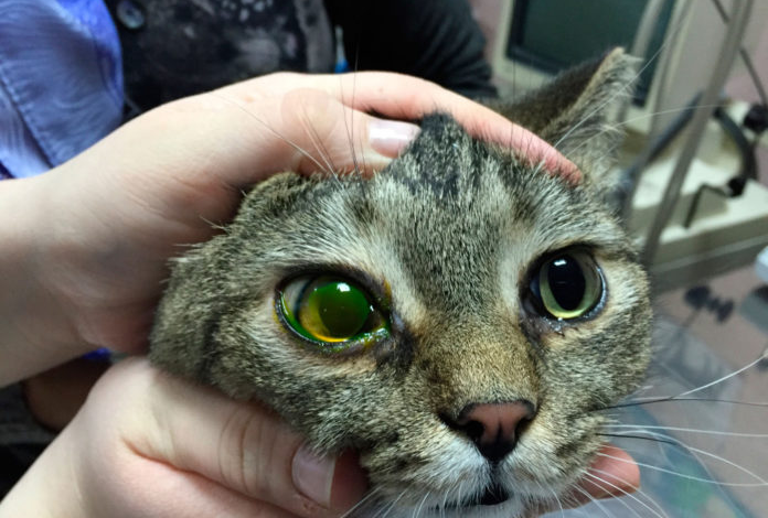
- The characteristic appearance of the cornea is one cloudy and swollen.
Seeing the ulcer itself at home is possible only in advanced cases with a perforated ulcer.
Based on etiological characteristics, three types of ulcers are distinguished:
- Purulent – occurs when there is a bacterial infection with abundant purulent discharge from the eyeball.
- Perforated – a complicated course, with penetration into the deep layers of the cornea.
- Creeping – occurs when infected with staphylococcus or streptococcus, quickly occupies a vast territory.
You should not diagnose a corneal ulcer on your own. It is almost impossible to do this at home without special equipment. Therefore, when the first signs of eye disease appear, it is important to immediately contact a veterinarian in order to begin treatment in a timely manner and preserve your pet’s vision and the organ itself.
One of the simplest diagnostic methods is to instill a special fluorescent solution – fluorescein – into the conjunctival sac. After this, the condition of the cornea is assessed under the light of a slit lamp. In this case, defects on the surface, as well as the extent of the lesion, will be clearly visible. Also, this test can determine the depth of the ulcer, the condition of the cornea itself and the smallest erosions.
As additional research, to detect the causes of the disease and concomitant diseases and complications, the following is carried out:
- Schirmer test – performed to diagnose dry eye syndrome. The test is carried out using express strips that determine the amount of tear fluid.
- Measuring intraocular pressure using a non-contact method – using a computer that determines the response of the cornea to an air stream.
- Bacterioscopy is carried out from material taken from the ulcer itself. Helps to accurately identify the pathogen and prescribe adequate treatment.
A corneal ulcer is a serious disease that leads to dangerous complications and can lead to removal of the eye. Therefore, you should definitely consult a doctor to make an accurate diagnosis, since an incorrect interpretation of symptoms can lead to an incorrect diagnosis and, accordingly, irrational treatment, which leads to a worsening of the condition.
Therapy depends on the causes that caused the disease and the pathogenic flora. During treatment, you should limit your pet’s physical activity and good, nutritious food. To avoid self-injury to the eye, a special collar is used.
Ulcers caused by bacteria require powerful antibiotic therapy. They are dripped locally, 6 times during the day. I usually use drops with a broad spectrum of action – ciprovet, Vigamox, iris, chloramphenicol, terramycin. To ensure long-term contact with the antibiotic (most often at night), ointments are used that are placed in the conjunctival sac – streptomycin.
Conservative therapy is suitable for a small defect that resolves without melting the cornea. In case of extensive damage, surgical intervention is possible. To do this, the bottom and edges of the ulcer are cleaned of necrotic tissue and the eye is covered with a special apron, which is formed from the third eyelid. It is also possible to simply stitch the edges of the eyelids.
After the operation, I prescribe systemic antibiotics – fluoroquinolones, penicillins, antibacterial drops into the conjunctival sac. The stitches are removed after 2 weeks. At the site of the ulcer, a scar should form, manifested as fibrosis of the cornea.
For viral cases, antiviral drops are used – trifluridine, idoxuridine. Prescribed daily until clinical signs disappear every 4 hours. Then the dosage is gradually reduced, continuing the course for 2 weeks. It is also advisable to use immunostimulating drugs.
If the cause is a foreign body or eyelashes, then they are removed immediately, even before treatment begins, in order to remove the provoking factor.
Under no circumstances should you use hormonal anti-inflammatory ointments and drops. They can lead to the spread of inflammation.
A modern surgical method for treating extensive ulcers is the transplantation of an artificial corneal graft.
- Timely treatment of inflammatory eye diseases and viral infections.
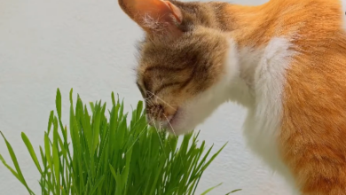
- Make sure that your pet does not walk through tall grass and bushes, and also control fights with street animals.
- Timely vaccination.
- Elimination of eye defects – eversion of the eyelid, third eyelid, eyelashes. And also trim off too long hair around the orbit.
How does a corneal ulcer occur in a cat and how dangerous is it?
The inflammatory process of the cornea is one of the most common and severe ophthalmological pathologies in domestic animals. An ulcer on an organ is accompanied by an inflammatory and necrotic process of epithelial tissue and stroma. Keratitis is dangerous due to the development of complete blindness and even loss of the eyeball. In addition to drug treatment, in many cases the pet will need surgical intervention.
Veterinary ophthalmologists, based on many years of practice, divide the causes of the development of ulcerative lesions of the cornea in domestic cats into non-infectious and infectious. Non-infectious factors that provoke the disease include:
 Mechanical damage to the cornea, eye injuries . Most often, cats belonging to brachycephalic breeds (Persian, exotic, Himalayan) are exposed to injuries to the delicate transparent membrane of the eye.
The peculiar structure of the skull and the convexity of the eye sockets in such animals predispose them to mechanical damage compared to other breeds.
Brachycephalics are also characterized by insufficient production of tear fluid (zophthalmos and lagophthalmos), xerosis of the central zone of corneal tissues, and structural features of the stroma. Together, these anatomical and physiological features lead to the frequent development of ophthalmological problems in animals.
- Foreign body . A sliver, twig, hard blade of grass, or metal shavings that get into the eye are a common cause of the development of ulcerative keratitis in a domestic cat.
- The cause of a corneal ulcer is often entropion – turning in of the eyelids . Ophthalmological pathology develops as a result of weakening of the ligamentous apparatus of the eyelid, as a result of which it tucks inside the eye. Wool and eyelashes, in contact with the delicate cornea, cause mechanical irritation and inflammation. Improper eyelash growth can also lead to a similar phenomenon.

A) Entropion of both eyes; B) Lagophthalmos
- Burn of the cornea of the eye by chemical products (acids, alkalis, etc.).
- Dry eye syndrome . Decreased tear production for any reason often leads to the development of ulcerative keratitis in domestic cats. Exophthalmos and lagophthalmos are the main causes of xerotic corneal ulcers in cats.
- Various types of conjunctivitis can develop into keratoconjunctivitis, and then into ulcerative keratitis.
- Damage to the trigeminal nerve is the cause of the development of neurogenic pathology.
- Disturbances in the innervation or blood supply to the tissues of the eye .
In addition to non-infectious causes, numerous factors of the disease are infectious agents. Viruses, bacteria, pathogenic fungi, rickettsia, chlamydia and other microorganisms, when they come into contact directly with the cornea or with the flow of lymph and blood from internal organs, lead to the development of an infectious process. The most common viruses that cause eye pathology are herpes virus and rhinotracheitis.
In the infection of corneal tissues by bacterial microflora, the leading place is occupied by staphylococci and streptococci. Chlamydia leads to an inflammatory process in the cornea, usually with generalized chlamydia infection in cats. Fungi most often provoke ulcerative keratitis when the animal’s immune system is suppressed.
What is a corneal ulcer?
A corneal ulcer in animals is a crater-shaped defect of the cornea caused by necrosis of its epithelium and stroma. Corneal ulcers in dogs and cats are among the most serious diseases and require immediate treatment by a veterinary ophthalmologist.

deep corneal ulcer that developed after an eye injury and improperly prescribed treatment.
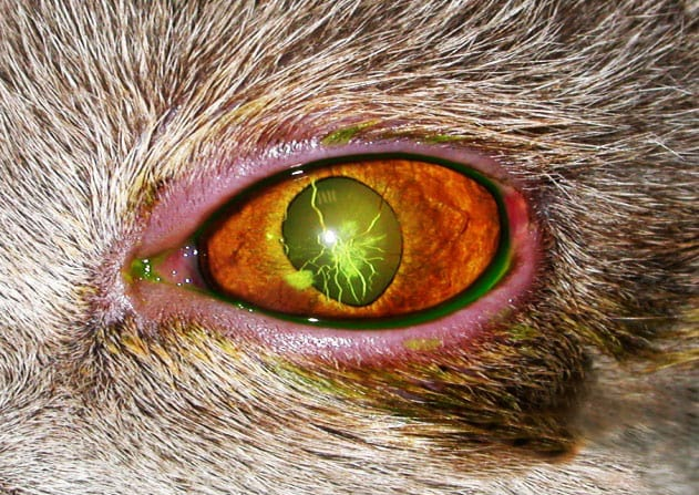
The danger of ulcers is due to their destructive effect on tissue, lightning-fast perforation of the eyeball, the development of purulent melting of all internal structures of the eye (panophthalmitis) and subsequent removal of the eyeball.

complication of corneal ulcer, purulent panophthalmitis. This eye needs to be removed urgently to save the healthy eye.
Other common severe complications of corneal ulcers are the development of secondary glaucoma and uveitis.
What is the mechanism of corneal ulcer formation?
Normally, the cornea in animals consists of four layers:
The outermost layer is the stratified epithelium. It protects the eye from infection getting inside. Unfortunately, the epithelium is damaged very quickly and then pathogenic microorganisms easily penetrate the corneal tissue.
Under the epithelium is the thickest membrane, the corneal stroma, which acts as a framework. The stroma consists of many transparent collagen plates located parallel to each other.
Behind the stroma there is a thin but very strong internal limiting membrane. It is the main protective barrier against all bacteria, viruses, foreign bodies and other damaging substances.
The innermost layer – the endothelium – is lined with only one layer of cells. The endothelium acts as an intraocular pump and does not recover when damaged.